In this section
Electron Microscopy Center
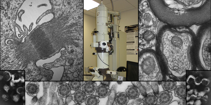
The Electron Microscopy Center (EMC) is a core facility located in room L.118C on the lower level of the Basic Sciences Building that houses a JEOL Transmission Electron Microscope (TEM). The TEM and equipment for sample preparation/processing is available on a cost-recovery basis to serve the diverse research interests of our students and faculty at Rosalind Franklin University of Medicine and Science. Arrangements for the preparation of your biological specimens can be made through the EMC Director.
Equipment and Facilities
JEOL JEM-1230, 120kV Transmission Electron Microscope (TEM) equipped with a high-resolution Hamamatsu ORCA-HR CCD Camera (2624 x 2400 pixel) with AMT Imaging Software, a Lab6 (long-life) filament , and a 5-axis motorized goniometer with position memory functions.
Special Functions Include:
- Accelerating voltages from 40-120kV
- Image Orientation System allows for electronic image rotation
- Minimum Dose System allows for beam-sensitive samples
- Through Focus allows for automation of focal series to optimize focus
- Separate investigator accounts store user-specific operating parameters
- CD/DVD data storage
Other ancillary equipment to support and facilitate specimen preparation includes: two Leica ultramicrotomes for thin-sectioning of specimens, a Leica rotary microtome for accommodating larger plastic embedded specimens (such as JB4 resin) for high-resolution optical microscopy, a Vibratome microtome for pre-slicing of whole specimens (such as brain), a Pelco easiGlow Glow Discharge System and a Biowave high-tech microwave oven. An on-site image analysis area includes: a Nikon Eclipse light microscope with a Nikon DS digital camera system, and a centrally located PC workstation.